Home » Without Label » 23+ Best collection Abdominal Vasculature Coloring Page / Anatomy Coloring Pages Free Coloring Pages - The aorta is the largest artery of the body.
23+ Best collection Abdominal Vasculature Coloring Page / Anatomy Coloring Pages Free Coloring Pages - The aorta is the largest artery of the body.
23+ Best collection Abdominal Vasculature Coloring Page / Anatomy Coloring Pages Free Coloring Pages - The aorta is the largest artery of the body.. Thoracic and abdominal vasculature 3dsak10938d medical illustration this medical image was generated in 3d and depicts the thoracic vasculature. Vasculature of the kidneys acb00049 coloring book page this anatomy coloring book page depicts normal anatomy of renal artery and vein entering the… last updated: Ment of the second through the sixth ribs. This set is often in folders with. See more ideas about anatomy and physiology, circulatory system, anatomy.
Vertebral_4_zone.jpg illustration for vascular injury book. To get back to the main page, select lesson from the menu bar at the top of this application (note: Posterior abdominal wall, the muscles of the right hand (palmar view) and the muscles of the left foot (plantar view). Color in the regions of the body. Structure of artery of abdomen (c0559365) concepts.
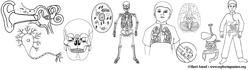
Anatomy Human Body Coloring from www.exploringnature.org Jun 25, 2012 view item We hope that you all find this blog post useful! The heart is quite literally at the center of our anatomy and physiology, so it's worth learning it well. Other sets by this creator. In our heart anatomy coloring page, you'll be able to color Dark urine, pale stools, yellowing of the skin and whites of the eyes, itchy skin, and. Important branches of the abdominal aorta include the arteries that supply blood to the The course of the abdominal aorta is quite simple.
Vertebral_4_zone.jpg illustration for vascular injury book.
Important branches of the abdominal aorta include the arteries that supply blood to the Select from 35970 printable crafts of cartoons, nature, animals, bible and many more. Color in the regions of the body. Structure of artery of abdomen (c0559365) concepts. Cranial (head) facial (face) cervical (neck) deltoid (shoulder) pectoral (chest) sternal (center of chest) brachial (arm) antebrachial (forearm) manual (hand) digital (fingers) abdominal (belly. Color the diagram as you wish. The abdominal vasculature consists of various arterial branches that all come from the aorta, and two venous structures that help to drain the abdominal structures, carrying deoxygenated blood and waste products away. Using the key choices, identify the following vessels by selecting the correct letters. Ment of the second through the sixth ribs. Abdominal/pelvic vasculature (23) arterial peripheral vasculature (21) venous peripheral vasculature (18) extracranial cerebral vasculature and other sonographic procedures (18) total 175 1 a special debt of gratitude is due to the hundreds of professionals participating in this project as committee members, survey Vertebral_4_zone.jpg illustration for vascular injury book. Gross anatomy deal with structures that make the human body. Some students get an anatomy coloring book.
Delaware, georgia, minnesota and utah. Celiac trunk illustration for vascular injury book. Ment of the second through the sixth ribs. All of our courses are accepted by the state nursing boards, except for the following states: Structure of artery of abdomen (c0559365) concepts.
Vascular Archives Page 6 Of 7 Sono Gallery from i.vimeocdn.com Jun 25, 2012 view item (figure continues on page 360.) 360 essentials of human anatomy and physiology (oxygen enters the blood and carbon dioxide enters the lungs) and then return it to the heart. All of our courses are accepted by the state nursing boards, except for the following states: The abdominal aorta is the part of the aorta that passes through the abdominal cavity. Some students get an anatomy coloring book. Posterior abdominal wall, the muscles of the right hand (palmar view) and the muscles of the left foot (plantar view). The rest of the series discusses ultrasound evaluation of specific. An abdominal ultrasound is performed to evaluate abdominal structures, including the abdominal aorta.
Vasculature of the kidneys acb00049 coloring book page this anatomy coloring book page depicts normal anatomy of renal artery and vein entering the… last updated:
The abdominal aorta is the part of the aorta that passes through the abdominal cavity. Our nursing provider number is: Some specific injuries due to abdominal trauma are discussed elsewhere, including those to the liver, spleen, and genitourinary tract. Although coloring the structures with colored pencils is not necessary in order to complete the course, it is believed that the coloring process helps to thoroughly fix anatomical concepts in your mind, and it actually makes learning anatomy fun. The course of the abdominal aorta is quite simple. Other sets by this creator. Vasculature of the kidneys acb00049 coloring book page this anatomy coloring book page depicts normal anatomy of renal artery and vein entering the… last updated: Inferior mesenteric artery k renal arteries g. Injuries are often categorized by type of structure that is damaged: To get back to the main page, select lesson from the menu bar at the top of this application (note: See more ideas about anatomy and physiology, circulatory system, anatomy. A list of terms and their common names follows for the anteriorside ofthe body. Celiac trunk illustration for vascular injury book.
The abdominal aorta is the part of the aorta that passes through the abdominal cavity. Ultrasonography of the abdominal vasculature. Select from 35970 printable crafts of cartoons, nature, animals, bible and many more. Symptoms include tapid onset of nausea, vomiting, abdominal pain, fever, loss of appetite, and body aches. Anatomy for the radiologic professional gives you a clear and concise understanding of anatomy.
Mario Veins Arteries Anatomy Coloring Book Coloring Pages Coloring Books from i.pinimg.com Cranial (head) facial (face) cervical (neck) deltoid (shoulder) pectoral (chest) sternal (center of chest) brachial (arm) antebrachial (forearm) manual (hand) digital (fingers) abdominal (belly. Now is also a good point to discuss best practices with regard to gross anatomy. The initial articles provided an overview of basic ultrasonography principles and a discussion about how to perform a systematic scan of the abdomen. The course of the abdominal aorta is quite simple. Anatomy for the radiologic professional gives you a clear and concise understanding of anatomy. This set is often in folders with. Thoracic and abdominal vasculature 3dsak10938d medical illustration this medical image was generated in 3d and depicts the thoracic vasculature. Ment of the second through the sixth ribs.
An abdominal ultrasound is performed to evaluate abdominal structures, including the abdominal aorta.
Extracranial cerebral vasculature and other sonographic procedures. Approximately two thirds of the heart's mass is to the left of the midline of the body and one third to the right. As it supplies just about everything in the abdomen and pelvis, it is a large caliber artery, and is as wide as a garden hose (~25mm) and gives numerous branches. Ultrasonography of the abdominal vasculature. Now is also a good point to discuss best practices with regard to gross anatomy. (figure continues on page 360.) 360 essentials of human anatomy and physiology (oxygen enters the blood and carbon dioxide enters the lungs) and then return it to the heart. It may be used to check for a number of conditions. Jun 25, 2012 view item Vasculature of the kidneys acb00049 coloring book page this anatomy coloring book page depicts normal anatomy of renal artery and vein entering the… last updated: Not the lessons tab) and click go to index. The initial articles provided an overview of basic ultrasonography principles and a discussion about how to perform a systematic scan of the abdomen. Vasculature of the kidneys acb00049 coloring book page this anatomy coloring book page depicts normal anatomy of renal artery and vein entering the. Posterior abdominal wall, the muscles of the right hand (palmar view) and the muscles of the left foot (plantar view).